Traditional Cornea Transplant
Understanding Cornea Transplants
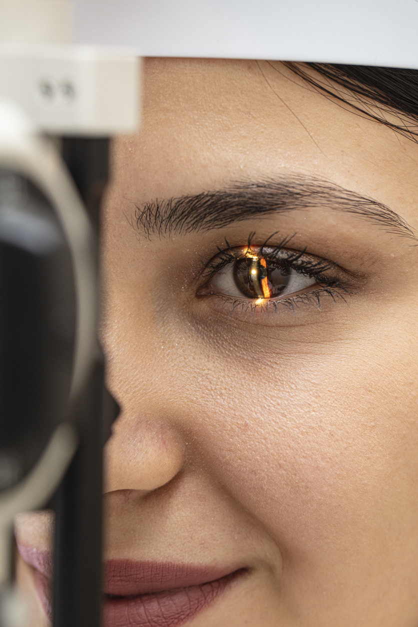
The cornea is the very front surface of the eye or the outer surface of the eye and plays a major role in focusing how you see images. If the cornea becomes weak or damaged, serious visual problems may arise. Treating damaged or irregular corneas is something that is routinely done at Eye Consultants of Atlanta. A damaged cornea requires special attention by our specially trained cornea eye surgeons. Often we initially treat the cornea with medication. If the issue with the cornea cannot be corrected with medications, eyeglasses or contact lenses, a corneal transplant or other procedure may be required.

How Does Damage to the Cornea Occur?
Damage to the cornea may arise from various conditions such as hereditary issues, chemical burns, blunt object trauma, and viral or bacterial infections. Conditions that may require a patient to seek a cornea transplant include clouding of the cornea, keratoconus, Fuchs’ dystrophy, irregular corneal surface tissue growths, or corneal scarring due to infection or trauma.
What Is a Corneal Transplant?
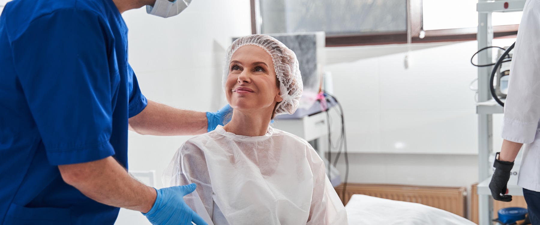
A corneal transplant is the removal of diseased corneal tissue with replacement of healthy tissue from an organ donor. There are several types of transplants, and the ideal surgery will be recommended by your corneal expert at Eye Consultants of Atlanta.

When to Have a Corneal Transplant
Your corneal expert at Eye Consultants of Atlanta will help you decide if and when it’s time for surgery. A common indication for corneal transplant is keratoconus. This condition is discussed in more detail below, and there are exciting new advancements for treating this condition. Specialty contact lenses may delay or eliminate the need for surgery.
For example, one study found that 69% of keratoconus patients that were referred for surgical intervention could instead be successfully fit with custom specialty contact lenses. . The Contact Lens Department at Eye Consultants of Atlanta can perform these custom fits. Thus, prior to transplant every effort should be made to optimally fit the patient with contact lenses, especially if there is not significant corneal scarring affecting vision. It is important to realize that a transplant does not necessarily guarantee freedom from contact lenses, as they are often required for resulting refractive errors and astigmatism.