Macular Degeneration

What Is Macular Degeneration?
Age-related macular degeneration (AMD) is the deterioration of the central retina, the macula. The macula is a small, specialized region of the retina and is responsible for central vision. Intact central vision is vital for reading, driving, and recognizing faces. Macular degeneration is a disease process that damages the central vision. It’s one of the leading causes of decreased vision in the United States in patients older than 50 years and the number one cause of blindness in Americans older than 65 years. Many older people develop mild forms of macular degeneration as a part of the natural aging process.
There are different kinds of macular problems, but macular degeneration is the most common macular disorder. Macular degeneration may manifest itself through symptoms such as blurriness, dark areas or distortion in central vision, and at end-stage, a permanent loss of central vision. Macular degeneration usually does not affect one’s peripheral vision. For example, with advanced macular degeneration, people can generally see the outline of a television, yet they may not be able to see the text of the TV screen.
Macular Degeneration Facts
- Those at risk – Individuals older than 60, but macular degeneration can appear as early as age 40. Macular degeneration is the most common cause of severe vision loss among people older than 65, and, as life expectancy increases, the disease is becoming more prevalent.
- The causes of AMD – There is no conclusive proof as to what causes macular degeneration. However, heredity plays a part, and other potential links include UV light exposure, poor vascular status, smoking, and malnutrition.
- The symptoms of AMD – The typical symptoms are distorted vision (typically straight lines or objects appear wavy or crooked), a dark spot in the central vision, and blurring. Later there is loss of central sight.
- What can be done to prevent AMD – Although there is no hard evidence as to how to prevent macular degeneration, there are various steps that may lower risks: proper nutrition, intake of daily vitamins, protection from UV sunlight, smoking cessation, and following nutrition guidelines found in the AREDS 2 (A.R.E.D.S.2) (Age- Related Eye Disease Study 2).
As mentioned, macular degeneration rarely occurs in people younger than 50 years and most commonly occurs in people older than 60 years. Macular degeneration is categorized into two major subsets,”wet” and”dry.”

Dry Form of Macular Degeneration
“Dry” signs of AMD include the accumulation of drusen (small yellow-white lumps) on the undersurface of the retina, loss of cells in the macula, and deterioration of the pigmented cells under the retina. Dry degeneration comprises 80% to 95% of macular degeneration. Mild forms of dry AMD cause little or no visual effects, but severe dry AMD may lead to blindness. There are currently new treatments for a subset of dry macular degeneration, but the results have not been overly promising. There is constant active research in this field. We expect to have better treatments for dry AMD in the coming years. Many studies have shown a benefit for the intake of dietary micronutrients. The AREDS 2 analysis has been shown to be more beneficial than AREDS 1, so we recommend the updated formula. Consumption of spinach or other green leafy vegetables, naturally colored foods rich in lutein and zeaxanthine (carrots, squash, bell peppers), and omega 3 fatty acids may help delay AMD as well. Smoking cessation is a key step, also.
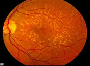


Mild Dry Macular Degeneration
Wet Form of AMD
The ”wet” form of AMD is also known as neovascular or exudative. Between 5% and 20% of all people with AMD have this form, and they may experience a more rapid loss of vision. In the wet form of AMD, new blood vessels develop beneath the retina in a layer of tissue called the choroid and grow into the macula. This process is called choroidal neovascularization, and the vascular network is called a choroidal neovascular membrane. Generally, the new blood vessels develop at the outer edge of the macula and extend inward toward the center of the macula. These abnormal vessels leak serum and blood. These fluids accumulate under the retina and separate the retina from the underlying layer, creating a localized retinal detachment. The retinal cells in the affected area do not function normally. This phenomenon causes central vision to become distorted, straight lines become wavy or crooked, and, eventually, a blind spot usually develops in the central vision. When advanced, wet et AMD can cause scarring of the central retina with an associated loss of central vision. As with dry macular degeneration, peripheral vision is rarely affected.
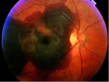
Severe wet macular degeneration showing blood accumulated below the retina
Symptoms of Macular Degeneration
Symptoms of AMD include distorted vision, a dark spot seen centrally, and blurred vision. Later, there is a loss of central vision. Central vision loss affects one’s ability to read, drive, and recognize facial features. Preservation of peripheral vision permits patients to see large objects and to retain ambulatory vision.
 (National Eye Institute photo)
The visual effects of end-stage macular degeneration with central vision obscured


How Is Macular Degeneration Diagnosed?
Your ophthalmologist uses a variety of examination and testing modalities to diagnose and assess macular degeneration. Eye Consultants of Atlanta has the latest and most advanced forms of analysis to help improve patients’ outcomes.
How Is Macular Degeneration Treated?
As mentioned, there is no known treatment for the ”dry” form of AMD, but multivitamins and certain dietary elements, AREDS 2, helps reduce the risk of progression. Smoking cessation is key, as well as leading a healthy lifestyle. In the coming years we anticipate the development of a medication that will help treat dry AMD. A low-vision evaluation at Atlanta’s Center for the Visually Impaired may offer visual benefits in the form of magnifying lenses, specialized lights, telescopic or prism glasses, and closed-circuit TV. Patients with the dry type of AMD may develop abnormal blood vessels and progress to the”wet” type of AMD.
Wet type of AMD has enjoyed several treatment breakthroughs in the last few years. Anti-VEGF (vascular endothelial growth factors) compounds are used to stop the growth of abnormal blood vessels. These medicines are administered on a regular basis and have provided hope for a previously hopeless disease.
Once Patients have received treatment for AMD, they must continue to have regular examinations of the retina so that any additional growth of new vessels can be detected and treated. New symptoms of visual distortion require prompt evaluation. As with dry AMD, a low-vision evaluation at Atlanta’s Center for the Visually Impaired may offer visual benefits in the form of magnifying lenses, specialized lights, telescopic or prism glasses, and closed circuit TV.
Anti-VEGF Compounds
This class of medications was originally created as cancer drugs to inhibit tumor growth. Anti-VEGF medications are now used to treat wet macular degeneration, and they work by inhibiting growth of abnormal blood vessels in the back of the eye.
These medications are intended to prevent further vision loss, and, in some cases, vision may improve with the use of anti-VEGF compounds
Anti-VEGF compounds are administered as intravitreal injections, i.e., the medication is placed directly into the vitreous of the eye. A careful sterile technique is used to administer the medication. Medication treatments are given at regular intervals for a period of time, and your eye physician will evaluate you every visit and modify the schedule accordingly. Complications from the treatment are extremely rare, and the sight- preserving benefits outweighs the risks.
Side effects of intravitreal injections may include:
- Serious eye infection that may include eye pain, light sensitivity, vision changes
- Increased eye pressure
- Retinal detachment
- Vitreous floaters
Is There a Way to Detect Retinal Changes in the Early Stages?
Early detection of macular changes is vital for proper treatment of macular degeneration. If you are at risk for developing macular degeneration, it is wise to check the vision in each eye daily. A self-administered test using an Amsler grid is a simple and quick way to check for sight changes. Call your ophthalmologist immediately if you notice changes on the grid.
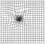
Abnormal Amsler grid
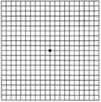
Normal Amsler grid
Instructions for the Amsler Grid Home Test
- Wear your reading glasses.
- Hold the Amsler Grid at a normal reading distance.
- Cover one eye.
- Look at the dot in the center of the grid.
- Note the appearance of the lines and squares. Assess for sight changes.
- Test the other eye in the same manner.
Understanding Your Results
- All of the lines should be straight and the squares of a uniform size
- If you note any changes in the appearance of the grid, such as distortion, blurring, discoloration, dark or missing areas of the grid, or any other changes, call your eye doctor immediately. Do not wait to see if the changes will clear up. Timely treatment is vital to safeguarding your vision.
The Prevention of Macular Degeneration
If you have a family history of macular degeneration or are a cigarette smoker, you may choose to make some lifestyle changes that could help lower your risk for macular degeneration. Listed below are a few tips for macular degeneration prevention.
- Regular exercise
- Weight under control
- Control of blood pressure
- Limiting UV exposure/wear quality sunglasses
- Obtain regular eye exams after the age of 40
- Do not smoke
- Follow AREDS2 nutrition guidelines
The information contained on this page is intended for educational purposes. Any patient with potential retina or eye disease or macular degeneration should consult an Atlanta ophthalmologist before making a judgment on their condition.
For more information on the retina in Atlanta, please contact us today to set up a consultation!